Thursday 19 November 2015

A Brief IntroductionTo: Imaging Flow Cytometers


We get a huge amount of data from our flow cytometers.  A guaranteed way to impress visitors unfamiliar with the technique is to start talking about the number of parameters we can measure simultaneously and the speed at which we can analyse millions of cells. But there are of course limitations.  Traditional flow cytometry tells us very little about cell morphology or cell-to-cell interactions and although we can find out which combination of fluorochromes is on, or in, each cell, we have no idea if the structures they are labelling are close together and interacting or entirely separated. All these things could be looked at by microscopy instead but that means sacrificing cell number so results are less robust, especially where rare events are concerned. To try to get around these problems we introduced  imaging cytometry into the lab a few years ago. We now also look after a second for one of our research groups.


What are they?

Essentially an imaging cytometer is the hybrid offspring of a flow cytometer and a microscope. It can analyse up to 12 fluorescent parameters (10 if using brightfield 9 if using both brightfield and scatter), giving you the same kind of information you’d get from any of our other bench top machines but with the addition of images of the cells. It does this using a CCD camera with a time delay integration (TDI) mode which tracks the cells and can give light and darkfield images as well as images of each fluorescent channel on up to 5000 events per second. The imaging cytometers aren’t as fast and don’t have as many fluorescent parameters as our top end benchtop machines but for many of our users the addition of the cell images more than makes up for that and sometimes dots on a screen just aren’t as convincing as actually seeing your cells. 


Our machines

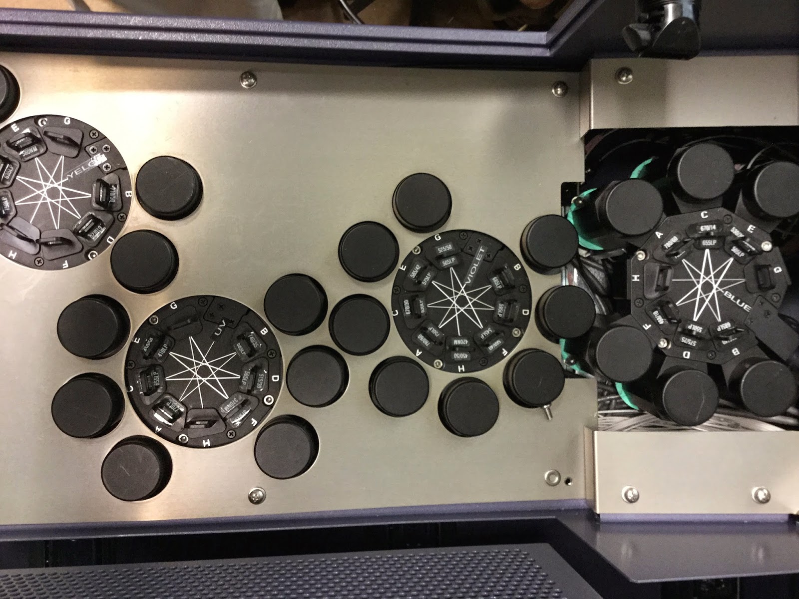
We have two imaging flow cytometers in the Crick Flow Lab. The Amnis Flowsight is a compact benchtop machine with 20x magnification and 9-12 fluorescent parameters that are detected by the camera, rather than by the PMTs used in conventional flow cytometers. It has blue (488nm), yellow (561nm), Red (642nm) and violet (405nm) lasers. Its big brother, the ImageStream X MKII, also has the 9-12 fluorescent parameters but comes with more powerful lasers and 20x or 60x magnification. The trade off of course is the price, with the ImageStream costing more and taking up more precious bench space. It also can’t run at full speed when using the 60x magnification. 


Using them

Actually running samples on the Imaging Cytometers is fairly straightforward, especially if you’re already familiar with conventional flow cytometers. Because a camera, rather than individual PMTs, detects the fluorescent parameters you can’t adjust the voltage on each parameter. Instead you need to tweak the laser power to alter the signal and this alters all the other fluorescent signals being analysed off that laser. This may mean you need to put more effort into fluorochrome selection and the concentration used in your sample. Something that works on a Fortessa might not automatically transfer to the ImageStream. My first attempt at cell cycle staining on the image stream just produced 10 channels full of PI with the channel I was interested in totally saturated and unable to tell me anything. But with a little bit of thought and sample prep good multicolour staining can be achieved.
The trickier part is analysing the data. Just like with any image analysis system it is possible to derive many metrics based on the images themselves as well as the localisation and spread of fluorescence. This does mean that to get the most out this technology, you need people to devote a good deal of time to the analysis and we have found, when training users, that analysis has to be tailored to their specific projects. 


Our work

Although imaging flow cytometry is a relatively new technique, we have done all sorts of things with it in the last few years. Amongst the applications where we have found imaging essential have been looking at cell morphology and cell size, cell division, co-localisation of signals, cell death, DNA damage, and small point fluorescence amongst other things.  


Sometimes with quite surprising results. Here are a few of the papers that came out of this work:
Mobilisation of Ca2+ in T Cells responding to different Stimuli
Asymmetric Cell Division
More Asymmetric Cell Division


Kirsty
 

Thursday 15 October 2015

Flow Lab FAQs - no.1: Tandems


We've set ourself a new task for our monthly lab meetings - take one of the questions we're often asked by our users and answer it in less than five minutes, with no slides. Although most of it is stuff that we all know already, it's always good to have a refresher and it provides an opertunity to discuss how we explain things to users to make sure they get acurate and consistent information.

We'll be sharing some of these Flow Lab FAQs on this blog, first up:


What are Tandem Conjugates and what are their Pros and cons?


What are they?

Tandems are essentially two fluorochromes chemically coupled together:


With single fluorochromes we use a laser to excite the molecule to a higher excitation state, then look at the energy given off as the molecule returns to its ground state.


Tandem conjugates use FRET (Förster, or fluorescence, Energy Resonance Transfer) so that the energy emitted by the first fluorochrome (A, the donor dye) is passed to the second (B, the acceptor dye). B is then excited to a higher state before dropping back down and emitting energy that we can detect.


The Benefits:


The difference between the excitation wavelength and the emission wavelength of a fluorochrome is known as the Stokes Shift. The size of this shift is limited in single fluorochromes but can be greatly increased by using a tandem. This means that with limited excitation sources (lasers) you can make use of more of the visual spectrum.


The Problems


No matter how efficient the process of making a tandem, there will always be some residual fluorescence from the donor dye. If a sample is stained with PE and PE-Cy7 antibodies it will be impossible to tell the difference between that and the PE signal from the tandem.  Even worse, this changes over time. Fortunately we can do something about this as we know it will happen. We also need to take more care with the storage of tandem dyes as their emission spectra will change over time.


Precautions To Take When Working with Tandem Conjugates

Controls
Controls
Controls


·      When using a tandem, ALWAYS have a single colour control

·      Always use the exact same antibody in your control as in your panel.
o   Different batches of tandems will always be slightly different so it is essential for correct      compensation that the exact same batch is used for the controls and the sample.
o   If your population is too rare or expressed at too low a level for a good control, use beads instead of cells

·      Always check the single control in the channels for its component parts to see donor emission.

·      Tandems tend to be more photosensitive than single fluorochromes so need to be protected from light during storage and during and after staining.

·      Tandems are also more prone to fixation issues as fixation will always alter the chemical structure of the dyes so make sure the controls and samples are treated in the same way.

·      When you get a new batch of a tandem, it’s a good idea to run it and look at the spillover, e.g. PE-Cy7 spillover in the PE channel. Then follow this over time. If the spillover starts to increase the efficiency of the FRET in the tandem is probably becoming less.

·      The name of most tandems makes it obvious what they are but that isn’t the case for some newer fluorochromes. Eg. Brilliant Violet BV 605 is a tandem of BV421 and Cy3.5 (all BV dyes above 605 are tandems). All the same precautions should be taken with these less obvious tandems.

Kirsty


Sunday 13 September 2015

Summer update


It's been a busy time for us here at the Crick Flow Cytometry Facility. Not only have we been trying to align the services we provide over our Mill Hill and Lincoln's Inn Fields sites but we have also been active in the cytometry community. In addition, we have been preparing for the big move to the Crick which is now planned for early 2016. As you can imagine moving around 80 Research Labs and 14 Science Technology platforms is not a trivial task. However our last visit to the site showed that the space allocated for the Flow Cytometry Facility is almost ready and is looking good - we are excited about the move!

Since we last posted, a number of the Lab managed to attend CYTO 2015, the annual meeting of ISAC, the International Society for the Advancement of Cytometry. This is the place to be to get the latest updates on hardware, software and reagents but more importantly it is a chance to network with colleagues. This year, CYTO was held in Glasgow at the Scottish Exhibition and Conference Centre. There was a good deal of excitement about the new multiparameter analyser from Propel Labs, the Yeti, and Mass Cytometry in the form of the Helios from Fluidigm continues to make progress but it seemed that the theme of the meeting was the use of microfluidics either for sorting or analysis and this may well be an area that we need to expand into in the coming years.

Prior to the meeting, a two-day hands-on course was provided to local students. This was organised by ISAC's  Live Education Delivery Task Force and flowcytometryUK. Around 50 delegates were able to see the latest equipment which was provided by several manufacturers and be able to pick the brains of 14 world-recognised experts in the field.

The opening lecture by Nobel Laureate, Eric Betzig

The pre-Congress course. Crick involvement in the DNA module!

Glasgow University


One of the most important aspects of a meeting like CYTO is that is gives a Forum for discussion and we were involved with the organisation of a workshop looking at how Core Facilities can make sure they are appropriately acknowledged in publications and how they can even take control of their own publication destiny. A similar workshop was also run recently at the IV Core Management meeting in Lausanne, Switzerland. The major points that came out of these workshops was that cores of all kinds, not just the Flow Cytometry ones, face similar issues - how can we keep track of publications that result from work done in the Lab, how can we publish the technological work that is done in our facilities and how can we ensure that we are seen not just as a 'service'.

Finally, we recently completed two 'bonding days' for our Labs at Mill Hill and Lincoln's Inn Fields where we managed to get (nearly) everyone together - keep an eye on the Blog for further team photos!


The Crick Flow Cytometry Team
Derek

Monday 18 May 2015

Attune NxT Demo

Recently we had the Attune NxT cytometer in the lab for a demo. The Attune can have up to 14 parameters (plus forward and side scatter) from up to four lasers. The demo model had blue, yellow, red and violet lasers. Optical filters can easily be swapped. You'd expect all that from any new flow cytometer but the Attune does have a unique feature - acoustic focusing.


As any Flow pros reading this will know, most other flow cytometers use hydrodynamic focusing to produce a very narrow core of sample, pushing the cells past the lasers in an orderly line. The problem with this is that if you have a large volume of sample to get through the cytometer it can take a very long time. Turning up the flow rate will speed things along but means that that narrow sample core gets a lot wider. This gives the cells a bit of room to move around so they won’t all pass through the laser interrogation points in a uniform way with the result that the data collected will have broader CVs.

The Attune’s acoustic focusing is designed to get around this problem. It uses sound waves which act like ripples in the stream, pushing all of the cells into the center, even when a large volume is being run. On it’s fastest setting the Attune can analyse 1000ul per minute, with a maximum event rate of 35,000 events per second. 


Other benefits of the Attune are that it’s pretty small (just as well given how little bench space there is left in our LIF Lab at the moment), and all fluidics are on board. It’s a syringe driven system so it doesn’t get through huge volumes of sheath fluid and it should be possible to get absolute counts from it too, although this function wasn’t available with the version of the software we had.

The rest of the software seems quite user friendly especially if you’re used to recent versions of Mircrosoft Windows and Office, but with that in mind, it would have been really nice to have a touchscreen. The big buttons on the monitor look like you should be able to tap on them (something I did automatically several times).

Time was limited for the demo and we didn't get to do as much as we would have liked on it. But we did have a bit of a play and the speed is certainly impressive with very dilute samples. A sample that ran at a rather tedious 24 evts/sec on the lowest flow rate zipped along at 800 evts/sec on the 1000ul/ml setting. As promised the CV remains pretty good even at the highest flow rate.


PI stained DNA samples run at the lowest and highest speeds.


The automated compensation is also very easy and speedy, although I did have very good controls (of course). The software was a little buggy in places, on a few occasions nothing showed up on the Scatter plot although the evts/sec indicator showed the sample was running and it can be slow to clear/refresh the plots which is a nuisance when trying to set up the voltages. Hopefully these issues will be fixed in the newer version of the software. 

The other issue was with carry over between samples. After running a particularly concentrated batch of beads it took a deep clean to get rid of them, the standard "rinse" function wasn't enough. So this could be an issue for those with sticky cells.

Overall the Attune NxT seems pretty good and would be especially suited to those who want to run large volume samples in order to find rare events or just to avoid the inevitable cell loss that comes with processing and spinning down a sample.  

Kirsty


Tuesday 28 April 2015

Introducing The Fortessa X30


Our Mill Hill site recently took delivery of the first Fortessa X30 in the country. As promised, here are a few more details...

This is the flagship flow cytometer from BD Biosciences which allows for the analysis of 27 fluorophores as well as forward and side scatter. Ours has 5 lasers: a 355nm UV, a 405nm violet, a 488nm blue, a 561nm yellow/green and a 640nm red. The signals generated from each laser line are detected by an octagon of PMTs allowing for up to 8 fluorophores per laser. Currently the cytometer is configured for 6 UV parameters, 8 violet, 5 blue, 5 yellow green and 3 red. As with the standard Fortessas, the optical filters can easily be changed, opening up a huge range of potential fluorophores and this number will only increase as new fluorophores are developed. The X30 hardware also has the potential for further development with the addition of more PMTs up to a maximum of 60 further enhancing the range of fluorescent markers available.


Graham putting the X30 through its paces


But there is a lot we can do with what we currently have. Even for those who don't need a huge number of fluorochromes, the X30 and our two new X20's will be a big help. The number of different lasers and detectors available means a wider choice of fluorochromes so we can really optimise what we use for each marker and how they will combine, potentially with less compensation and data spread too. But these machines will have the biggest impact on our users who are already working with 13 or 14 colour experiments and keen to expand still further. With the X30 on site and the recent addition of brilliant UV and brilliant violet dyes to the market, huge multicolour experiments are on their way to the Crick!

We'll report back with more on this once we start really putting it through it's paces and getting some results. In the mean time, here's a little look inside...

Phil and Kirsty.


Octagons and a lot of PMTS
Red laser octagon


Wednesday 1 April 2015

Welcome

Welcome to the Crick Flow Blog.

Today sees the official launch of the Francis Crick Institute and with it, the formation of the Crick Flow Cytometry Facility.

The existing flow cytometry / FACS labs at the former Cancer Research UK, London Research Institute (LRI) and the MRC's National Institute for Medical Research (NIMR) will be joining forces to become the biggest (and we hope best) flow cytometry core facility in Europe.


Staff currently based at the LRI.
Left to Right- Derek (lab head), Joana, Kirsty, Sukhveer, Marwa and Debi. Also (not in the picture) Carl.


For the next few months we will be operating across both existing locations but our current users should only notice a  couple of changes: Firstly, we will be moving over to a new booking system which should go live very soon and secondly everyone will be seeing some new faces as the staff begin to work at both sites.

We've already begun to take delivery of some exciting new equipment including two twenty parameter Fortessa X20's and a custom built Fortessa X30, the first of its kind in the UK. More on those very soon...


   Inside the new Fortessa x30 at the NIMR site


The new X20 at our LRI site - just coming out of the wrappers


We are scheduled to start physically moving into our new lab in December of this year. We will go in three phases, the last of which is scheduled for May 2016.  We got a sneak peek at our new space earlier in the month, it's all coming together really well and our rooms have been carefully situated close to most of the labs the will use us the most.


Today also sees the launch of this blog, along with our twitter account and YouTube channel, boldly taking the flow cytometry core facility into the world of social media. Our plan is that these will keep the flow cytometry world, as well as our users, up to date as we embark on the exciting challenge of bringing two well established labs together and starting a new era with the Francis Crick Institute.

Make sure you also subscribe to our Twitter feed for updates on the lab and our YouTube Channel for some handy how to videos, we'll be adding more to that and this blog in the coming months.


Kirsty