Monday 19 December 2016

The Crick Flow Lab - We're In!


It’s been years in the planning and taken months of packing, unpacking and calling in what feels like every flow cytometry engineer in the UK, but the Francis Crick Institute and its Flow Cytometry Facility are finally up and running.


The building was officially opened last month by Her Majesty the Queen, accompanied by the Duke of Edinburgh, Prince Andrew and a host of other VIP guests. But we’d already been in the building for weeks getting our equipment back up and running after it was moved from our “legacy” sites in Mill Hill and Lincoln’s Inn Fields.

Sir Paul Nurse Introduces The Queen to some Crick Scientists
photo by, Fiona Hanson


The move happened in three stages to ensure that we were able to offer a service to users at the new site as soon as they arrived while still having machines on the old sites for those who moved later. This meant we were amoungst the first in and the last out on all three sites. By the time of the last move the old labs, which used to be so busy, were starting to feel eerily deserted, but they also had more bench space than ever before!


The old Lincoln's Inn Fields Lab and it's last remaining Fortessa


Inevitably things didn’t go entirely smoothly. That was never likely with a move of this size and complexity and there were delays getting our Containment Level 2 hoods ready, a few things got broken in transit and we discovered some very very small lab coats. But with a lot of work, help from all those engineers and the wonderful Crick staff, most of our equipment is now open for business.

The Crick has been designed to make full use of Science Technology Platforms (STPs) such as ours. Immunology labs and others with the greatest need for Flow Cytometry facilities have been clustered close to us. Anyone having to come from further afield (over 12 floors - the building covers 1,000,000 square feet) can follow designated routes to ensure samples don’t have to be carried through the many informal meeting and breakout areas.

The Crick Flow Facility itself is made up of five rooms. We have a prep lab with our own tissue culture hood, incubator etc. The old labs had very little bench space so we are now much better equipped to process users samples and stain cells to test out new techniques. We then have two analyser rooms. One of those houses our two imaging cytometers, a MacsQuant VVB and a Fortessa X20. The latter will soon be in a hood that will allow us to do Containment Level 2 analysis without the need to fix cells first.


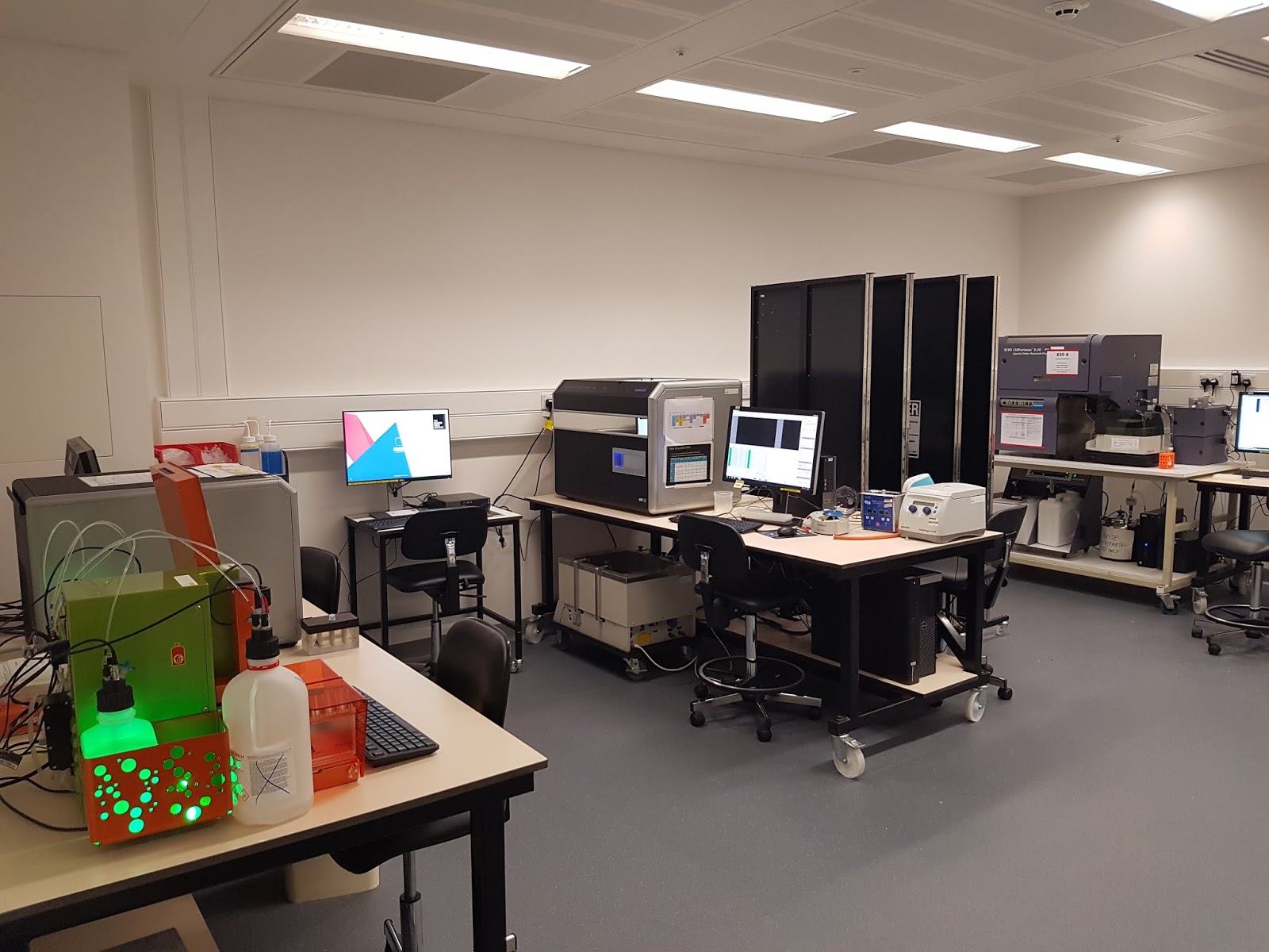
Our second analyser room currently contains six five laser Fortessas, three LSRII’s, a FACSVerse and three computers loaded with a range of analysis software.


Our sorting facility is divided between the Containment Level 2 and Containment Level 1 rooms. In the former, we have an Aria III, an XDP and two Influxes in hoods and in the latter there are two more XDPs plus two Aria Fusions for the non hazardous samples. All of these machines will be operated exclusively by members of the flow cytometry facility. Howevere, we also have an Avalon sorter which gives users the option of doing simpler sorts for themselves at times when the lab isn’t staffed or everyone is booked up on the more complex sorters.


The challenge of bringing together staff and users from two quite different institutions was perhaps almost as great as moving all our instruments. In the months leading up to the physical move we began swapping staff between sites to help all the lab members get used to working together and to introduce ourselves to our new users. The flow cytometry lab now has 12 staff and the Crick is home to around 1250 researchers - so that’s potentially quite a lot of people to train and sort cells for.

We are starting to get used to the new lab now but still have lots to do, lots of new people to meet and plenty of plans for the future. We’ll try to update this blog as it all happens and will also be adding more to our YouTube channel and twitter account.


A rare photo of the entire Crick Flow lab staff in one place
(L-R) Joana, Bhavik, Graham, Phil, Carl, Kirsty, Damian, Florian, Wayne, Sukhveer, Debi and Derek

Kirsty

Sunday 11 September 2016

Conferences and a Lab move!

Well, it's been a busy few months with two major conferences and now the start of the move to the new Crick building. June saw the annual CYTO Conference this year held in the Washington State Convention Centre in Seattle which was also the venue in 2010 (but this time we were not hampered by Icelandic volcanos!). I like Seattle, it has a laid-back vibe but has everything you need. The conference itself is really a must-attend for those in the Flow Core business - this is where we get to see the companies newer products as well as hear about cutting-edge cytometric developments from leaders in the field. Plus it's a great chance to interact with like-minded people and socialise.

This year saw a number of new machines on the market - the new Helios from Fluidigm which brings mass-cytometry to a wider audience; the simple, almost self-service sorter, the FACS Melody from Becton Dickinson, and the promise of a new 6 laser CytoFlex from Beckman Coulter. All technologies that will make our lives in the core easier and will expand the capabilities of cytometric systems. The exhibition side of CYTO meetings is always interesting, plenty of people who are willing to talk enthusiastically about their products but generally without the hard sell; many thanks to ISAC's Organising Committee for their hard work once again in putting on the meeting.

Another important feature of CYTO is the more informal sessions; the tutorials where experts give up their time to explain to newcomers the intricacies of hardware, sofware or specialist tecniques; the workshops where facilitators direct discussion on a particular topic - especially relevant this year were several workshops devoted specifically to Core Facility Management or working; and the CYTO innovation session, which I found incredibly engaging.

Of course CYTO isn't CYTO without a major closing night celebration and this year we closed the conference with drinks and buffet at the Seattle aquarium (no fish were harmed during the course of this party!).


After barely a month's recovery, we headed to Leeds for flowcytometryUK 2016. It may be a smaller affair but it was a great success. Over 180 delegates and exhibitors enjoyed nearly three days of presentations, workshops (both scientific and commercial), posters and an exhibition. This was co-organised by myself and Rachael Walker from the Babraham Institute along with support from the Royal Microscopical Society. We managed to attract a great line-up of speakers - ISAC President Paul Wallace, flow gurus Paul Robinson and Howard Shapiro, one of the founders of flow Lee Herzenberg (who was presented with an RMS Honorary Fellowship at the meeting) plus some of the UK's rising cyometry starts Jim Brewer, Muzz Haniffa and Wolf Reik.

And if that wasn't enough at the beginning of August we began our move into the now fully-built Francis Crick Institute. This will be the subject of a future blog when we have completed the move (we are going over three phases so we can keep work going at our new building and our legacy sites) but as a taster, here are a couple of pictures.

Looking out of the building to the East

The view from the Office and write-up area

Since we moved in there is no need for the PPE!


Thursday 12 May 2016

Using Video Guides To Help Lab Users


Training new Lab users on all our benchtop analysers is a big part of our role in the Flow Cytometry facility. All new users, even if they have a lot of previous flow cytometry experience, will have one-to-one training with a member of our Lab. This could be as simple as going over the cleaning and data storage procedures for those who know everything else already. Or it could involve several sessions where we cover the principles of the technique and the entire process from experimental design to analysis and data presentation. Lab users will only be allowed full 24/7 access to the instruments once we are happy that they know what they are doing and, importantly, that they will look after our precious machines!

Inevitably though people forget things, and problems occur when we're not around to help. Over the years, emails have been sent out and our machines have accumulated reminder notices. The problem is though, people don’t always read the signs. If they have been there for a while, they just fade into the background. Also, it can be difficult to explain a physical process with just a few words and a couple of pictures. But any more than that and people may not bother to read it at all. What you really need is a moving, talking human in the Lab. But none of us is terribly keen on being here all night just in case someone forgets how to top up the sheath tank.

To try to solve this problem, we’ve created some short videos. The most recent one shows users how to set up and shut down the new HTS. The HTS lets users run samples straight from a 96 well plate and although they are common in a lot of labs including our Mill Hill site, they haven’t been used much here at Lincoln’s Inn Fields.

However, we recently fitted one to our Fortessa X20 and it’s been quite popular with users who don’t want to spend hours sat in front of a flow cytometer just to swap a tube over every five minutes. It's a pretty straightforward system to use and saves time when there are a lot of samples to analyse. But it does require the user to physically change over a few parts on the machine (and put them back again afterwards). So rather than writing a lengthy flat pack furniture style text description (insert tube A into port B …etc, etc.) Joana filmed me doing the swap, I uploaded the video to YouTube and then we made it available to our users:


Making the video guides on a smartphone (Samsung S5) was very quick and straightforward. The guides use a combination of still images and video, edited together on the phone and rather than talking through the instructions there are subtitles. So users don't need sound.

To make it easier to find the videos when they are needed I used free online tools to create shortened URL and QR codes. These are stuck to the HTS so anyone with a smartphone and a QR reading app can just point their phone at it and the video will pop up. It’s a tip I’d seen mentioned on Twitter for putting instructions on a communal coffee machine, something which is probably more complicated to use than the HTS (although messing up a macchiato isn’t usually as bad as ruining 96 samples).


The HTS video is easily accessed using a QR code and shortened URL on the machine


We also have a short video for all our analysers, reminding people how to change the sheath and waste tanks, plus a few troubleshooting tips. 


Of course the videos are no substitute for proper training by a real life flow expert, but realistically there are times when users who should perhaps know better, forget the basics when no one is around to help, especially if it’s a problem they don’t often encounter. So I don’t think we’ll be winning any Oscars but hopefully the video guides and easy access QR codes will help out our users when they have late night problems and save us from finding dried up cytometers or ruined experiments the next morning.


If we ever get a coffee machine that (and the accompanying video of course) might help too. 

Thursday 28 January 2016

Flow Lab FAQs - no.2: Antibody Titration


Our second Flow lab FAQ is something we probably wish we were asked about more often... 


Antibody Titration -  How And Why?

Titration allows the identification of the optimal concentration of a given reagent in a particular experiment. It's worth doing because sometimes, the concentration recommended by the company, or in a paper, is not the best to use in your assay.


Why?

Titration will help you to find the best signal-to-noise ratio for your staining and therefore give you the best chance of detecting a weak signal. It may also save you money if the optimal concentration is less than the manufacturer's recommendation:

All fluorescence measurements have associated background noise, which comes mainly from two sources. Some intracellular molecules, like NADH, fluoresce when excited at certain wavelengths (autofluorescence). The flow cytometer also has inherent noise coming from detectors and electronics. 


The background noise can increase if the concentration of the dye/antibody is too high, because it will start binding not only to the high-affinity epitopes it's supposed to target, but also to low affinity ones (non-specific binding). In practical terms, the median of the negative signal will increase causing a drop in the staining index. You can see this drop in the green arrow on the plot below.


How:

Stain a known number of cells with a range of different antibody concentrations. For example, if the recommended concentration is 10μM you can test 2.5, 5, 10 and 20μM. Then analyse them on your flow cytometer. It is very important that the staining conditions are kept the same in the titration and in the experiment (incubation time, temperature, fixation, etc), because the staining level is affected by experimental conditions. The volume of the reaction is also critical, try to keep this constant.

 Which concentration should I choose after the titration?
 
To select which concentration (or range of concentrations) is the best to use, calculate the STAIN INDEX for each tested value. The stain index is calculated as follows:  


Use FlowJo or any other cytometry analysis software to calculate the median of the positive and negative signals and the standard deviation of the negative signal.
 
The concentration with highest stain index should be selected for use on the experimental panel. In the graph above, the best range of concentrations is marked with a red box.


Frequently Asked Questions

1.    Should I use a viability dye in the titration?
If possible, yes. Dead cells usually have higher levels of autofluorescnce and can bind antibodies non-specifically

2.    Can I use compensation beads for the titration?
No! With titration we are looking for the specific binding of an antibody to antigen. Compensation beads work by binding the light chains and the antibody NOT the specific binding site.

3.    My marker is really rare. Can I test the same fluorochrome targeting a more abundant marker?
No! We are trying to establish the antibody binding NOT the level of fluorescence. If the marker is rare in your cells, try to find a cell line where it is highly expressed; manufacturers datasheets will often say which cells have been used to test the antibody.

Remember - It is important to keep the same staining conditions for the titration and for the final experiment. 

Joana

Wednesday 6 January 2016

2016 - A Big Year!


Not just because it has an extra day, but 2016 will be big for the Crick Flow Cytometry Facility. At some point in the middle of this year we will begin moving into the brand new Francis Crick Institute. Situated in the heart of London, next to St Pancras station and the British Library, the Crick will be one of one of the largest biomedical research Institutes in the world - you can read about that here.

For the past 18 months we have been preparing for the merger of the old London Research Institute and National Institute for Medical Research Flow Labs by aligning common practices and cross-training of staff and we are, we think, ready for the move. Because both Labs are large, we plan to move in phases so that we can keep operations running on both sites until all researchers make the move to the new building. It is logistically difficult but we like a challenge!

The Francis Crick Institute, London

In amongst all that, we need to make sure that we keep up with developments in the field - Twitter is a great source of information so via @citometria we were led to this article about using sound to sort cells. We will keep an eye (or maybe an ear) on that.

2015 was a good year for meetings - there was the first UK CYTO meeting for almost 30 years in Glasgow and then in November we had the biennial "Advances in Cytometry" meeting in London plus the usual mix of local flow meetings in London, Cambridge, Aberdeen, Nottingham and more. This year, we have CYTO in Seattle which is always a great place to meet up with old friends but also to see the new innovations in the field, I am always amazed at the breadth of cytometric applications and the passion with which people advocate the technology. But for those of you who can't make that, there is flowcytometryUK2016 which will be held in Leeds in July - planning is underway for that and there will be considerable Crick Flow Lab involvement - more on that later in the year.

We also hope to use this Blog to help promote cytometry education by putting up bite-sized answers to questions we often get from users, our Flow Lab FAQs - so bookmark this page or follow us on Twitter of course!

Derek

Thursday 19 November 2015

A Brief IntroductionTo: Imaging Flow Cytometers


We get a huge amount of data from our flow cytometers.  A guaranteed way to impress visitors unfamiliar with the technique is to start talking about the number of parameters we can measure simultaneously and the speed at which we can analyse millions of cells. But there are of course limitations.  Traditional flow cytometry tells us very little about cell morphology or cell-to-cell interactions and although we can find out which combination of fluorochromes is on, or in, each cell, we have no idea if the structures they are labelling are close together and interacting or entirely separated. All these things could be looked at by microscopy instead but that means sacrificing cell number so results are less robust, especially where rare events are concerned. To try to get around these problems we introduced  imaging cytometry into the lab a few years ago. We now also look after a second for one of our research groups.


What are they?

Essentially an imaging cytometer is the hybrid offspring of a flow cytometer and a microscope. It can analyse up to 12 fluorescent parameters (10 if using brightfield 9 if using both brightfield and scatter), giving you the same kind of information you’d get from any of our other bench top machines but with the addition of images of the cells. It does this using a CCD camera with a time delay integration (TDI) mode which tracks the cells and can give light and darkfield images as well as images of each fluorescent channel on up to 5000 events per second. The imaging cytometers aren’t as fast and don’t have as many fluorescent parameters as our top end benchtop machines but for many of our users the addition of the cell images more than makes up for that and sometimes dots on a screen just aren’t as convincing as actually seeing your cells. 


Our machines

We have two imaging flow cytometers in the Crick Flow Lab. The Amnis Flowsight is a compact benchtop machine with 20x magnification and 9-12 fluorescent parameters that are detected by the camera, rather than by the PMTs used in conventional flow cytometers. It has blue (488nm), yellow (561nm), Red (642nm) and violet (405nm) lasers. Its big brother, the ImageStream X MKII, also has the 9-12 fluorescent parameters but comes with more powerful lasers and 20x or 60x magnification. The trade off of course is the price, with the ImageStream costing more and taking up more precious bench space. It also can’t run at full speed when using the 60x magnification. 


Using them

Actually running samples on the Imaging Cytometers is fairly straightforward, especially if you’re already familiar with conventional flow cytometers. Because a camera, rather than individual PMTs, detects the fluorescent parameters you can’t adjust the voltage on each parameter. Instead you need to tweak the laser power to alter the signal and this alters all the other fluorescent signals being analysed off that laser. This may mean you need to put more effort into fluorochrome selection and the concentration used in your sample. Something that works on a Fortessa might not automatically transfer to the ImageStream. My first attempt at cell cycle staining on the image stream just produced 10 channels full of PI with the channel I was interested in totally saturated and unable to tell me anything. But with a little bit of thought and sample prep good multicolour staining can be achieved.
The trickier part is analysing the data. Just like with any image analysis system it is possible to derive many metrics based on the images themselves as well as the localisation and spread of fluorescence. This does mean that to get the most out this technology, you need people to devote a good deal of time to the analysis and we have found, when training users, that analysis has to be tailored to their specific projects. 


Our work

Although imaging flow cytometry is a relatively new technique, we have done all sorts of things with it in the last few years. Amongst the applications where we have found imaging essential have been looking at cell morphology and cell size, cell division, co-localisation of signals, cell death, DNA damage, and small point fluorescence amongst other things.  


Sometimes with quite surprising results. Here are a few of the papers that came out of this work:
Mobilisation of Ca2+ in T Cells responding to different Stimuli
Asymmetric Cell Division
More Asymmetric Cell Division


Kirsty
 

Thursday 15 October 2015

Flow Lab FAQs - no.1: Tandems


We've set ourself a new task for our monthly lab meetings - take one of the questions we're often asked by our users and answer it in less than five minutes, with no slides. Although most of it is stuff that we all know already, it's always good to have a refresher and it provides an opertunity to discuss how we explain things to users to make sure they get acurate and consistent information.

We'll be sharing some of these Flow Lab FAQs on this blog, first up:


What are Tandem Conjugates and what are their Pros and cons?


What are they?

Tandems are essentially two fluorochromes chemically coupled together:


With single fluorochromes we use a laser to excite the molecule to a higher excitation state, then look at the energy given off as the molecule returns to its ground state.


Tandem conjugates use FRET (Förster, or fluorescence, Energy Resonance Transfer) so that the energy emitted by the first fluorochrome (A, the donor dye) is passed to the second (B, the acceptor dye). B is then excited to a higher state before dropping back down and emitting energy that we can detect.


The Benefits:


The difference between the excitation wavelength and the emission wavelength of a fluorochrome is known as the Stokes Shift. The size of this shift is limited in single fluorochromes but can be greatly increased by using a tandem. This means that with limited excitation sources (lasers) you can make use of more of the visual spectrum.


The Problems


No matter how efficient the process of making a tandem, there will always be some residual fluorescence from the donor dye. If a sample is stained with PE and PE-Cy7 antibodies it will be impossible to tell the difference between that and the PE signal from the tandem.  Even worse, this changes over time. Fortunately we can do something about this as we know it will happen. We also need to take more care with the storage of tandem dyes as their emission spectra will change over time.


Precautions To Take When Working with Tandem Conjugates

Controls
Controls
Controls


·      When using a tandem, ALWAYS have a single colour control

·      Always use the exact same antibody in your control as in your panel.
o   Different batches of tandems will always be slightly different so it is essential for correct      compensation that the exact same batch is used for the controls and the sample.
o   If your population is too rare or expressed at too low a level for a good control, use beads instead of cells

·      Always check the single control in the channels for its component parts to see donor emission.

·      Tandems tend to be more photosensitive than single fluorochromes so need to be protected from light during storage and during and after staining.

·      Tandems are also more prone to fixation issues as fixation will always alter the chemical structure of the dyes so make sure the controls and samples are treated in the same way.

·      When you get a new batch of a tandem, it’s a good idea to run it and look at the spillover, e.g. PE-Cy7 spillover in the PE channel. Then follow this over time. If the spillover starts to increase the efficiency of the FRET in the tandem is probably becoming less.

·      The name of most tandems makes it obvious what they are but that isn’t the case for some newer fluorochromes. Eg. Brilliant Violet BV 605 is a tandem of BV421 and Cy3.5 (all BV dyes above 605 are tandems). All the same precautions should be taken with these less obvious tandems.

Kirsty